Medical Image Analysis – The Role of AI in Modern Medical Diagnostics

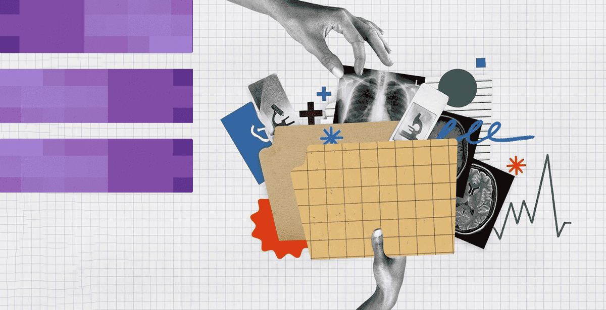
A radiologist reviews hundreds of scans a week. Fatigue is real, and small findings get missed. Medical image analysis exists to catch what tired eyes sometimes don’t, without replacing the expert making the final call.
The technology has moved fast. What started as pattern-matching on X-rays now spans full pathology slides, dermatology photos, and endoscopy video.
If you’re interested in learning how medical image analysis actually delivers value, and where the hype still outruns the evidence, read on.
What Medical Image Analysis Actually Does
Medical image analysis uses computer vision models to detect, segment, and classify features inside clinical images. That includes tumors on a CT scan, cell structures on a pathology slide, or hemorrhages on a retinal photo.
The core technique is pattern recognition at a scale no human can match. A model trained on millions of labeled images learns which pixel patterns correlate with disease. It then flags similar patterns in a new image within seconds.
That speed doesn’t replace the clinician. It changes their workload. Instead of scanning every image with equal attention, a radiologist can prioritize the cases a model flags as high-risk first.
Computer vision services build this kind of segmentation and detection pipeline. That work spans a range of industries beyond healthcare alone. The underlying techniques, like U-Net segmentation and convolutional neural networks, are the same ones powering clinical imaging tools.
Three components make up nearly every medical image analysis pipeline. Preprocessing normalizes scans across different equipment and settings. A detection or segmentation model then isolates the region of interest. A final classification step assigns a diagnostic label with a confidence score attached.
Each stage introduces its own failure modes. Poor preprocessing can wash out subtle findings before the model ever sees them. A well-engineered pipeline treats all three stages as equally critical, not just the model at the center.
AI Medical Imaging: From Pixels to Diagnostic Signal
AI medical imaging systems don’t just look at pixels. They’re trained to recognize the diagnostic signal buried inside noise, artifacts, and natural anatomical variation.
Getting this right requires enormous, carefully labeled training datasets. A model trained on scans from one hospital’s equipment can perform worse on scans from a different manufacturer’s machine. Domain shift is one of the hardest problems in the field.
Labeling quality matters as much as model architecture here. Every training image needs annotation from a qualified specialist, and disagreement between expert annotators is common even among humans. A model can only be as reliable as the ground truth it learned from.
One of the strongest pieces of real-world evidence comes from a nationwide mammography study. Published in Nature Medicine, the PRAIM study followed 463,094 women across twelve German screening sites and 119 radiologists.
The results were striking. AI-supported screening detected 6.7 cancers per 1,000 women screened, against 5.7 per 1,000 in the control group. That’s a 17.6% relative increase in detection, without a rise in unnecessary recalls.
Just as important, radiologists spent 43% less time reading exams the AI tagged as normal. Average reading time dropped to 39 seconds from 67 seconds on those cases. That’s the workload relief clinicians actually need.
Radiology AI in Practice
Radiology AI is the most mature branch of medical image analysis today, and for good reason. Radiology produces standardized, digital images at massive volume, which is exactly what deep learning models need to train well.
Mammography, chest X-rays, and CT lung screening now have FDA-cleared AI tools in active clinical use. These tools typically work as a second reader, flagging cases for radiologist review rather than issuing an independent diagnosis.
Adoption is accelerating industry-wide. Regulatory clearances for AI and machine learning-enabled devices have grown steadily each year. Radiology still accounts for the largest share of authorized tools.
That growth brings real scrutiny, too. A cleared device needs post-market monitoring. Regulators want confirmation it performs as well in daily practice as it did in the validation study.
Performance can drift as patient populations and imaging equipment change over time. A model validated on one hospital’s patient mix may see different accuracy at a hospital serving a different demographic entirely.
Clinical workflow integration is the other underrated factor in whether a radiology AI tool actually gets used. A tool that adds clicks or interrupts a radiologist’s existing reading pattern gets ignored. That’s true no matter how accurate it was in a lab setting.
A Precision Benchmark from Outside Healthcare
DPL hasn’t yet deployed a clinical radiology AI system, and we won’t claim otherwise. But one of our computer vision engagements shows the exact discipline that healthcare-grade image analysis demands.
DPL built the verification engine behind Hafiz, the Digital Quran project, an initiative to authenticate sacred text across digitized manuscripts. The system had to handle wildly different calligraphy styles, scan qualities, and OCR error patterns.
The engineering challenge mirrors medical imaging closely. Both require extracting reliable signal from highly variable source images. Both then verify that signal against a trusted reference, with near-zero tolerance for error.
The result was a 99% precision rate authenticating text across formats that would confuse a simpler OCR pipeline. That same rigor, applied to segmentation and classification instead of text verification, is what clinical-grade image analysis requires.
Pathology Image Analysis: A Different Set of Constraints
Pathology image analysis faces a problem radiology doesn’t: image size. A single digitized pathology slide can be gigapixels in resolution, far too large to feed into a model at once.
Models have to work in tiles, analyzing small regions of a slide and then reassembling findings into a coherent whole. Getting the tiling strategy wrong means missing small but clinically significant features.
Cell-level detail also matters more in pathology than in radiology. Distinguishing between benign and malignant cells often comes down to nuclear shape and texture differences invisible at a glance. That demands finer-grained models than typical radiology use cases.
Machine learning consulting work applies this same tiling and fine-grained classification approach. Other high-resolution domains, like satellite imagery and industrial inspection, face the identical architecture challenge pathology imaging demands.
Healthcare Image Recognition Beyond the Hospital
Healthcare image recognition isn’t limited to hospital radiology departments anymore. Dermatology apps now screen skin lesions from a smartphone photo. Ophthalmology clinics use retinal imaging to catch diabetic eye disease before symptoms appear.
Remote and rural care benefit most from this shift. A clinic without an on-site specialist can capture an image and get an AI-assisted second opinion within minutes. That cuts what used to be a long wait for a referral.
Consumer wearables are pushing healthcare image recognition even further into daily life. Continuous skin monitoring and wound-tracking apps now exist for chronic care patients managing conditions between clinical visits.
None of this replaces specialist judgment. It extends specialist reach into settings that previously had none at all. That reach is where the technology’s social value is highest.
Where AI in Healthcare Is Headed Next
AI in healthcare is moving from single-task models toward multimodal systems. These combine images with clinical notes, lab results, and patient history. A model that only sees a scan misses context a clinician would naturally use.
Federated learning is also gaining traction. Hospitals can train shared models without ever moving sensitive patient images outside their own infrastructure. That directly addresses privacy concerns that have slowed adoption for years.
Regulatory frameworks are catching up too. Agencies are moving toward frameworks for continuously learning models instead of one-time approvals. Real clinical models keep improving after deployment, and oversight needs to keep pace.
Building Medical Image Analysis Systems Responsibly
None of this works without rigorous validation before deployment. A model tested only on data resembling its training set will fail silently on real-world edge cases it never saw.
Bias testing across demographics is not optional. Models trained predominantly on one population have historically underperformed on others, a documented failure mode across multiple imaging specialties.
Post-deployment monitoring matters as much as pre-deployment validation. DPL’s MLOps services practice exists precisely to catch model drift before it silently degrades diagnostic accuracy in production.
Explainability rounds out the requirements list. A clinician needs to see which part of an image triggered a flag. A bare probability score without that visual evidence rarely earns clinical trust.
Heatmap and saliency-map techniques help close that gap. They highlight the exact pixels driving a model’s prediction. A radiologist can then quickly confirm or override the finding based on their own read of that region.
Documentation matters just as much as the model itself. Every version, every training dataset change, and every validation result needs a clear audit trail. Regulators and hospital review boards will ask for it eventually. Scrambling to reconstruct that history after the fact is a preventable failure.
The Path Forward
Medical image analysis won’t replace radiologists or pathologists. It will change what they spend their limited attention on. Routine reads move aside so expertise goes where it matters most.
Getting there takes more than a promising model demo. It takes validated data pipelines and honest performance reporting across patient subgroups. It takes monitoring that catches drift before it reaches a patient.
The organizations getting this right start with that engineering discipline first. Use the form below to connect with DPL’s AI engineering team and bring that same discipline to computer vision problems across industries, healthcare included.

Topics
Related Articles
-

AI Chatbot Development Services – How to Build Bots That Solve Problems, Not Frustrate Users
11-Sep-2026 Talha Saleem
-
AI Video Analytics Services – Smarter Surveillance, Monitoring, and Operational Intelligence
08-Sep-2026 Salman Naseer
-
Prompt Engineering Services – The Hidden Lever Behind Better LLM Performance
07-Sep-2026 Hazar Hayat